
Introduction
This module will teach you about second heart sound auscultation including these learning objectives:
- Understand how S2 is generated
- Differentiate S2 from S1
- How to use the respiratory cycle to find S2 splitting
- Identify the types of S2 splitting
Before beginning this course, you should complete the Normal and First Heart Sounds modules and be able to recognize the first heart sound.
Overview
The second heart sound (S2) is the result of heart vibrations transmitted to the chest wall when the aortic (A2) and pulmonic (P2) valves close. These valves close at the end of systole.
Single S2
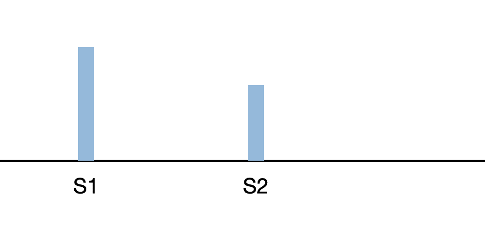
S2 is heard as a single sound when the aortic and pulmonic valves close simultaneously or nearly so. Because of a higher closing pressure, the aortic component is louder and closes slightly ahead of the pulmonic valve.
Physiologically Split S2
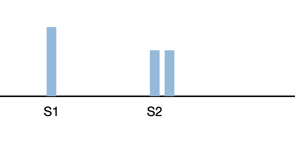
When the aortic and pulmonic components can be separately heard, this is known as a physiological split. This type of split is heard during inspiration. During expiration, a single S2 is heard. However, the degree of physiological splitting can decrease as adults age. Because the pulmonic sound component has less intensity than the aortic component, a split S2 is best heard at the pulmonic position.
Fixed Split S2
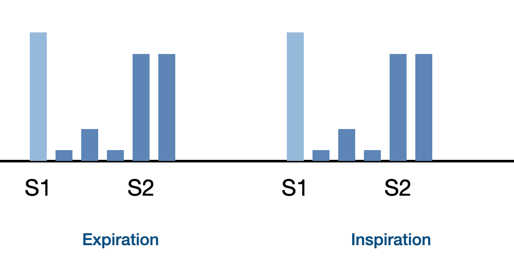
A discernible split that does not change during the respiratory cycle is a fixed split. Wide, fixed splits are associated with abnormal conditions such as atrial septal defect, acute pulmonary hypertension, and pulmonic stenosis. In the diagram, a fixed S2 split occurs along with a systolic murmur.
Start Second Heart Sounds Course
Second Heart Sounds Lessons
Physiologically Split S2
Closure of the aortic valve followed by the closing of the pulmonic valve generates the second heart sounds (S2). Careful observation of the intensity and splitting of the S2 can indicate the presence of several cardiac abnormalities.
In this recording, we present normal physiological S2 splitting. The splitting varies between zero and eighty milliseconds over the respiratory cycle. The S2 aortic component (S2A) precedes the S2 pulmonic component (S2P).
Physiologically Split S2 Lesson
Persistent Splitting S2
In this recording, we can observe S2 persistent splitting. Splitting varies between thirty milliseconds at peak expiration and sixty milliseconds at peak inspiration.
With normal physiologic splitting, the second heart sound is not split at peak expiration. But with persistent splitting, the S2 splits during inspiration and expiration. However, the amount of splitting is reduced in expiration.
This type of splitting is associated with pulmonary hypertension and right bundle branch block
Persistent Splitting S2 Lesson
Fixed Splitting S2
This lesson and recording presents fixed splitting of the second heart sound. The S2 split is unchanged through inspiration and expiration at about sixty milliseconds.
Fixed S2 splitting is associated with acute pulmonary hypertension, pulmonary stenosis, and pulmonary emboli. Fixed splitting of the second heart sound along with murmurs can indicate the presence of an atrial septal defect, a congenital heart defect.
The murmurs have been eliminated in this recording so that you can concentrate on the fixed splitting of the second heart sound. In a later module you will hear the fixed splitting of S2 along with murmurs of atrial septal defect.
Fixed Splitting, Increased Aortic Intensity S2
Listen to this recording of a second heart sound with fixed splitting and including an increased sound intensity of the aortic component. This condition is seen in some patients with chronic, severe hypertension; aortic pressure is significantly increased.
Our recording contains fixed splitting of S2 to make it easier to concentrate on the relative intensities of the aortic and pulmonic components. In the cardiac animation video, note the increased thickness of the left ventricular wall caused by essential hypertension.
Fixed Splitting, Increased Aortic Intensity S2 Lesson
Fixed Splitting, Decreased Aortic Intensity S2
Second heart sound (S2) with fixed splitting and an aortic component with decreased intensity is presented in this lesson. We are using a fixed splitting audio recording to help you more easily hear the difference in intensity between the aortic and pulmonic components of S2.
Fixed Splitting, Decreased Aortic Intensity S2 Lesson
Late Systolic Click and S2
Note that certain heart sound patterns can resemble S2 splitting. A late systolic click plus a single-second heart sound (S2) in one such condition. In this case, a late systolic click occurs immediately before the second heart sound. This incorrect leads to a finding of a split S2.
To correctly evaluate the sounds, carefully to the two sounds. If the first of the pair is of higher frequency and shorter duration than the second, it is a late systolic click followed by a single second heart sound.
Moving the stethoscope chestpiece to the pulmonic area is also a good technique for distinguishing a late systolic click from a split S2. The single S2 will be present and the late systolic click will diminish or disappear
Degeneration of the mitral valve leaflets causes these leaflets to make a "clicking" sound during late systole. In the video, you will see prolapse of the anterior lateral mitral valve leaflet which generates the late systolic click.
Late Systolic Click and S2 Lesson
Tumor Plop and S2
A single S2 followed by a tumor plop in diastole can also mimic a split second heart sound.
If you move the stethoscope chestpiece to the pulmonic position, the tumor plop diminishes or disappears. But if two distinct sounds at the pulmonic area are observed, then this is likely to be an S2 split.
Tumor plop and third heart sound gallop have the same timing and frequency. It is not possible to distinguish one from the other.
Opening Snap and S2
In this recording, listen for an opening snap that begins about 75 milliseconds after the commencement of S2. Such opening snaps in early in diastole and a single S2 can mimic a split second heart sound.
The source of an opening snap is thickened valve leaflets which produce a snapping sound when opening. The more severe the thickening the earlier in diastole the opening snap occurs.
Course Quiz
After completing all lessons in a course, a quiz becomes available. If the user successfully completes a quiz, results are saved to the user's dashboard and a certificate can be printed.
Reference Guide
For subscribers, we provide a comprehensive heart sounds and murmurs reference guide. For each abnormality, one or more sound recordings are available along with text, phonocardiogram and cardiac animation.Course Completion
Registered users can earn a certificate of achievement for this module by reading all content and then earning a passing score on this module's quiz.
Completed modules and related scores can be viewed on the dashboard.
Authors and Reviewers
-
Heart sounds by Dr. Jonathan Keroes, MD and David Lieberman, Developer, Virtual Cardiac Patient.
- Lung sounds provided by Diane Wrigley, PA
-
Heart sounds mentorship by W. Proctor Harvey, MD
- Reviewed by Dr. Barbara Erickson, PhD, RN, CCRN.
-
Last Update: 11/10/2022
Sources
-
Heart Sounds and Murmurs Across the Lifespan (with CD)
by Dr Barbara Ann Erickson
Publisher: Mosby
ISBN-10: 0323020453; ISBN-13: 978-0323020459 -
Heart Sounds and Murmurs: A Practical Guide with Audio CD-ROM 3rd Edition
Elsevier-Health Sciences Division
Barbara A. Erickson, PhD, RN, CCRN -
Heart and Lung Sounds Reference Guide
PracticalClinicalSkills.com -
Heart Sounds Made Easy with CD-ROM: (with CD-ROM) 2nd Edition
Anthony P. Salmon
ISBN-13: 978-0443069079 - NCBI Review of Heart Sounds and Murmurs: A Practical Guide
-
The Virtual Cardiac Patient: A Multimedia Guide to Heart Sounds And Murmurs
Jonathan Keroes, David Lieberman
Publisher: Lippincott Williams & Wilkin)
ISBN-10: 0781784425; ISBN-13: 978-0781784429 -
Ventricular Function Curves in the Exercising Dog
JONATHAN KEROES , ROGER R. ECKER , and ELLIOT RAPAPORT
Circulation Research, Vol. 25, No. 5 -
Electrocardiographic changes associated with ritodrine-induced maternal tachycardia and hypokalemia
American Journal of Obstetrics Gynecology, VOLUME 154, ISSUE 4, P921-923, APRIL 01, 1986
Susan K Hendricks, MD, Jonathan Keroes, MD, Michael Katz, MD -
The_Virtual_Cardiac_Patient_A_Multimedia_Guide_to_Heart_Sounds_and_Murmurs
A Multimedia Guide to Heart Sounds and Murmurs
January 2007 JAMA The Journal of the American Medical Association 297(2):217-218
DOI:10.1001/jama.297.2.217. M. Saleem Seyal, MD, Reviewer -
Clinical Heart Disease
W Proctor Harvey, MD
Laennec Publishing; 1st edition (January 1, 2009)