
Introduction
This course presents third vs fourth heart sounds. Before beginning this course, you should finish the Normal, First and Second Heart Sounds courses.
- Understand how S3 and S4 are generated
- Differentiate S3 and S4 from S1 and S2
- Identify the cadence patterns of S3 and S4 gallops
- Using the correct auscultation techniques
Overview
The third heart sound (S3) is a low-frequency, short vibration occurring in early diastole at the end of the rapid diastolic filling period of the right or left ventricle. S3 may be heard in normal children.
Third Heart Sound Gallop
In adults, hearing a third heart sound requires quiet. Ask the patient to exhale and hold their breath. Shift stethoscope bell around the apical and lower sternal area listening for a third heart sound gallop. The cadence is similar to 'Kentucky'.
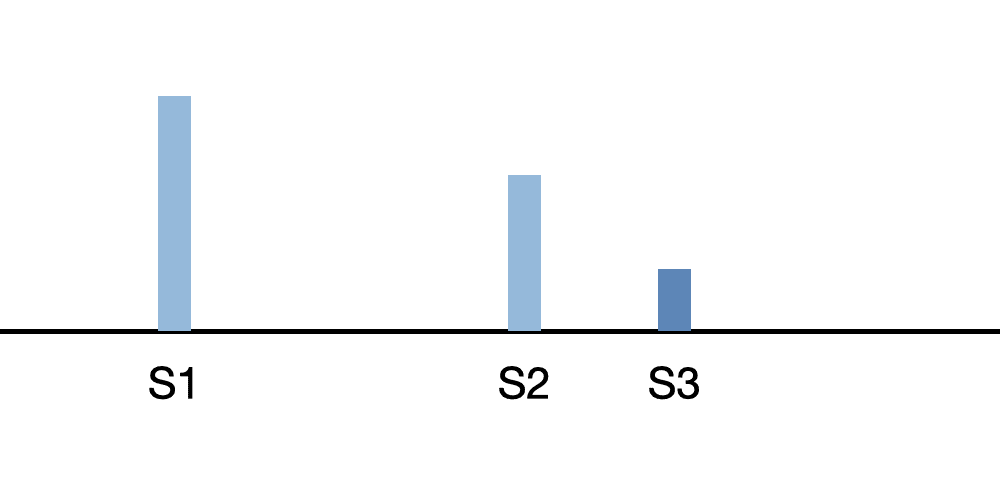
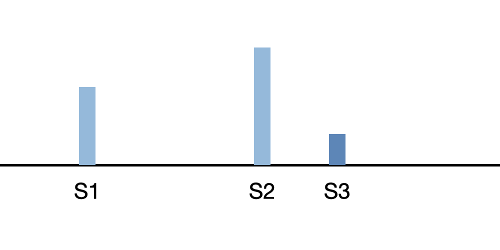
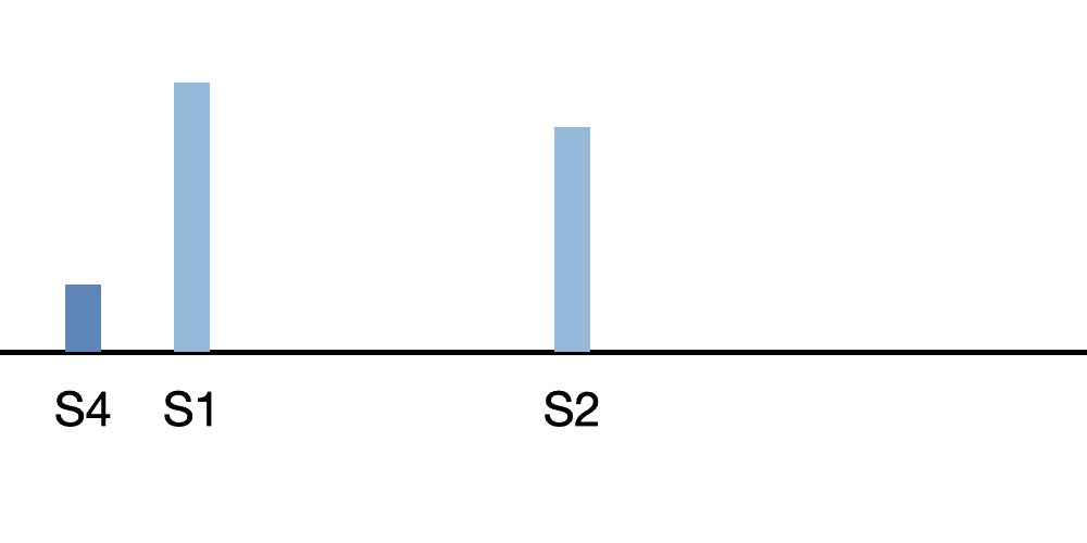
Sound Patterns
As an introduction, we present two sound patterns that illustrate S3 and S4 gallops. We will cover these in the lessons that follow.
Third Heart Sound - Heart Disease
In adults over the age of 40, S3 can indicate heart disease, except for patients that are athletes. As you will hear in the lessons, S3 is a low-pitched sound, allowing the observer to distinguish S3 from a split S2.
Fourth Heart Sound Gallop
The fourth heart sound (S4) is a low-pitched, late diastolic sound. So it occurs shortly before the first heart sound. The stethoscope bell should be used, pressing lightly against the chest wall at the lower left sternal border or subxiphoid area. The cadence is similar to 'Tennessee'. S4 will often indicate a heart abnormality.
Start Third and Fourth Heart Sounds Course
Lessons
Third Heart Sound Gallop
The third heart sound (S3) is a low-frequency sound best heard with the stethoscope bell, pressed lightly on the skin. A third heart sound occurs early in diastole. In the presence of S3, the first heart sound has diminished loudness. The second heart sound becomes louder.
Sudden deceleration of blood flow into the left ventricle from the left atrium causes the third heart sound. Observe the dilated, thin-walled left ventricle with generalized decreased contraction vigor in the cardiac animation video.
In young people and athletes, it is a normal phenomenon. In older people, S3 indicates congestive heart failure.
In addition to sound intensity, the first heart sound (S1) can be identified by its timing. At moderate heartbeat rates, the first heart sound follows the longer pause.
Third heart sounds are also called ventricular gallops or S3 gallops.
Third Heart Sound Gallop Lesson
Fourth Heart Sound Gallop
In the recording, note that the fourth heart sound (S4) occurs in late diastole just before S1. The S1 intensity is decreased, while S2 intensity is increased. The fourth heart sound is low-pitched. Use the stethoscope bell, pressing lightly on the chest wall.
The fourth heart sound is generated by increased left ventricle stiffness due to scar tissue formation. This condition could be due to coronary heart disease.
Essential hypertension and aortic stenosis can also lead to fourth heart sounds. The anatomy animation video provides an example of S4.
Fourth Heart Sound Gallop Lesson
Third and Fourth Heart Sound Gallop
Listen to the recording of a third and fourth heart sound gallop. Note that the third and fourth heart sounds are low frequency. S4 has a pitch somewhat lower than S3.
Patients with heart failure will often have a third heart sound. As the patient's condition improves, this S3 becomes a third and fourth heart sound gallop.
Third and Fourth Heart Sound Gallop Lesson
Summation Gallop at 120BPM
With high heart rates, in this case 120 BPM, the diastolic interval is shortened. Accordingly, S3 and S4 sounds begin to merge, producing a single loud heart sound.
The cardiac animation video presents an enlarged left ventricle with decreased left ventricular contractility and a minimally enlarged left atrium.
Course Quiz
After completing all lessons in a course, a quiz becomes available. If the user successfully completes a quiz, results are saved to the user's dashboard and a certificate can be printed.
Reference Guide
For subscribers, we provide a comprehensive heart sounds and murmurs reference guide. For each abnormality, one or more sound recordings are available along with text, phonocardiogram and cardiac animation.Course Completion
Registered users can earn a certificate of achievement for this module by reading all content and then earning a passing score on this module's quiz.
Completed modules and related scores can be viewed on the dashboard.
Authors and Reviewers
-
Heart sounds by Dr. Jonathan Keroes, MD and David Lieberman, Developer, Virtual Cardiac Patient.
- Lung sounds provided by Diane Wrigley, PA
-
Heart sounds mentorship by W. Proctor Harvey, MD
- Reviewed by Dr. Barbara Erickson, PhD, RN, CCRN.
-
Last Update: 11/10/2022
Sources
-
Heart Sounds and Murmurs Across the Lifespan (with CD)
by Dr Barbara Ann Erickson
Publisher: Mosby
ISBN-10: 0323020453; ISBN-13: 978-0323020459 -
Heart Sounds and Murmurs: A Practical Guide with Audio CD-ROM 3rd Edition
Elsevier-Health Sciences Division
Barbara A. Erickson, PhD, RN, CCRN -
Heart and Lung Sounds Reference Guide
PracticalClinicalSkills.com -
Heart Sounds Made Easy with CD-ROM: (with CD-ROM) 2nd Edition
Anthony P. Salmon
ISBN-13: 978-0443069079 - NCBI Review of Heart Sounds and Murmurs: A Practical Guide
-
The Virtual Cardiac Patient: A Multimedia Guide to Heart Sounds And Murmurs
Jonathan Keroes, David Lieberman
Publisher: Lippincott Williams & Wilkin)
ISBN-10: 0781784425; ISBN-13: 978-0781784429 -
Ventricular Function Curves in the Exercising Dog
JONATHAN KEROES , ROGER R. ECKER , and ELLIOT RAPAPORT
Circulation Research, Vol. 25, No. 5 -
Electrocardiographic changes associated with ritodrine-induced maternal tachycardia and hypokalemia
American Journal of Obstetrics Gynecology, VOLUME 154, ISSUE 4, P921-923, APRIL 01, 1986
Susan K Hendricks, MD, Jonathan Keroes, MD, Michael Katz, MD -
The_Virtual_Cardiac_Patient_A_Multimedia_Guide_to_Heart_Sounds_and_Murmurs
A Multimedia Guide to Heart Sounds and Murmurs
January 2007 JAMA The Journal of the American Medical Association 297(2):217-218
DOI:10.1001/jama.297.2.217. M. Saleem Seyal, MD, Reviewer -
Clinical Heart Disease
W Proctor Harvey, MD
Laennec Publishing; 1st edition (January 1, 2009)