
Introduction
This systolic murmurs module includes seven lessons. We provide a textual description, audio recording, dynamic waveform video, and a cardiac animation in each lesson. Optionally, a quiz can be taken to measure comprehension and listening skills. Users who have selected our Auscultation-Basics, Essentials or Advanced plans can print achievement certificates and view their progress and scores using our personalized dashboard.
- Differentiate systolic from diastolic murmurs
- Identify murmur timing
- Recognize key features of the murmur's sound
- Gain exposure to some abnormalities associated with these murmurs
Systolic murmurs are sustained sounds occurring between the first and second heart sounds. These murmurs can be normal in many patients, including infants, children. In adults, some systolic murmurs are normal, such as in pregnancy.
When auscultating, take note of these murmur attributes:
- Location
- Timing: early, mid, late or pansystolic
- Loudness and its pattern during systole
- Pitch
- Quality (harsh, blowing or musical)
- Murmur changes during the respiratory cycle
Sound Patterns
As an introduction, we present three sound patterns that illustrate systolic murmurs. We will cover these in the lessons that follow.
Crescendo Decrescendo
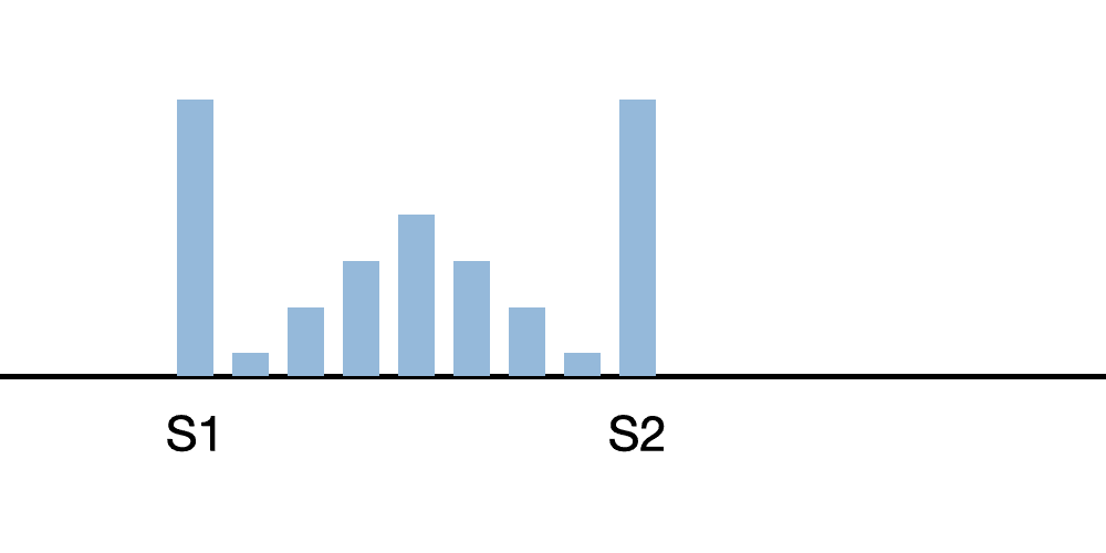
A common sound pattern for systolic murmurs is a crescendo/decrescendo. In this case, the murmur is moderate and S1, S2 are louder than the murmur.
Severe Aortic Stenosis
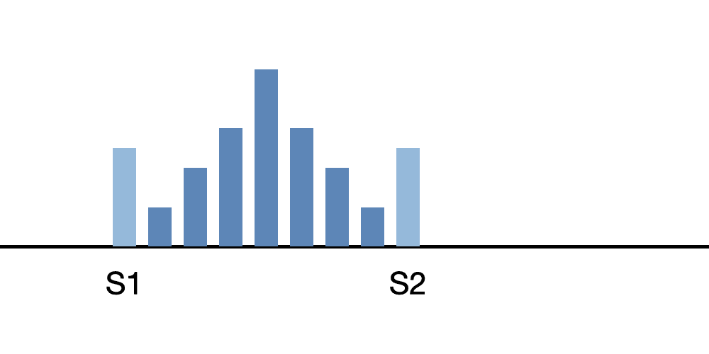
Here the systolic murmur is present in a patient with severe aortic stenosis.
Innocent Murmur
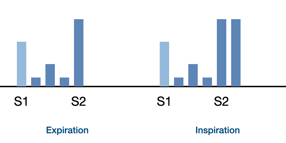
Here a young patient presents with an innocent murmur and S2 split.
Start Systolic Murmurs Course
Systolic Murmurs Lessons
The lessons in this course present several types of abnormal systolic murmurs, their attributes, and clinical correlations.
Innocent Murmur
This recording is of an innocent murmur. Innocent murmurs are generated by normal blood flow within the heart. Blood flow turbulence creates vibrations in the heart's tissues which are then transmitted to the chest wall.
Benign, innocent murmurs are very common in children. This type of murmur is associated with non-cardiac conditions such as pregnancy, hyperthyroidism, exercise, and anemia in adults. When these conditions are treated appropriately, the systolic murmur disappears.
Use the stethoscope bell or diaphragm, auscultating over the pulmonic area. Listen for increasing sound intensity with inspiration. Innocent murmurs usually appear in early systole with low-to-mid-range-frequencies of 100-250Hz. The murmur is fairly short in duration. In this recording S1 and S2 are normal. Diastole is silent.
Aortic Sclerosis Heart Sound
This aortic sclerosis murmur is fairly loud, heard early in systole. When viewing the waveform, notice the diamond-shaped appearance. Regular vibrations of this murmur provide a musical quality ("cooing").
It is caused by turbulent blood flow into the aorta. Diastole is silent and S1 and S2 are normal.
Mild Aortic Stenosis
Aortic stenosis has a murmur characterized by an aortic ejection click in early systole, followed by a diamond-shaped systolic murmur that ends mid-way through systole. The murmur has a mid-frequency pitch. As the condition worsens, the murmur frequency increases.
The first heart sound is normal. The second heart sound is physiologically split. The aortic component of the second heart sound is louder than normal.
The anatomy video shows thickened aortic valve leaflets with decreased movement. The left ventricle wall thickness is increased.
Severe Aortic Stenosis
In severe aortic stenosis, there is a diamond-shaped systolic murmur continuing through systole. The murmur is loud and higher pitched than a mild aortic stenosis murmur.
S1 is normal. S2 is louder than normal. In fact, you are hearing only the accentuated pulmonic component of S2 due to heart failure on the left side. The aortic ejection click heard in mild cases of valvular aortic stenosis is gone. A fourth heart sound can often be heard in late diastole. This is caused by the increased left ventricular wall thickness and stiffness.
Calcification of the aortic valve leaflets is a cause of this murmur. When viewing the cardiac animation, look for a greatly thickened left ventricular wall and the almost totally immobile aortic leaflets.
Mitral Regurgitation
A mitral regurgitation murmur is a rectangular murmur that continues throughout all systole. It has a mid-frequency pitch. S1 is normal. S2 is single. There is a third heart sound gallop in diastole.
The murmur is generated by turbulent blood flow through the incompetent mitral valve leaflets into the left atrium. Both the left ventricle and the left atrium are enlarged.
Mitral Valve Prolapse
The mitral valve prolapse murmur is medium pitched with a diamond shaped waveform. This murmur begins immediately after a mid systolic click and continues to the end of systole.
The intensity of the murmur increases and its starting point begins earlier in systole as left ventricular volume decreases (going from supine to standing). The intensity of the murmur and its starting position move later in systole as the volume increases (by raising the legs while in the supine position). The mid-systolic click also moves in tandem with the murmur.
On the cardiac animation video, observe that the murmur is caused by the prolapse of the posterior mitral valve leaflet. The murmur is illustrated by turbulent flow from the left ventricle into the left atrium.
Hypertrophic Cardiomyopathy
Hypertrophic cardiomyopathy associated murmur is an early peaking, harsh, diamond shaped, systolic murmur. It begins at the onset of systole and stops well before S2. A fourth heart sound gallop is also present in diastole. View the waveform to see these features.
Observe that S1 intensity has increased due to a hyperdynamic left ventricle. S2 is single.
On the anatomy video, observe that the contraction of the left ventricle is strong and occurs in a reduced amount of time. Anatomically, the septal wall is very much thicker than the rest of the ventricle, but this is not shown in the animation.
The strong contraction of the left ventricle causes the anterior leaflet to be sucked into the ventricle, blocking the flow into the aorta and causing an aortic murmur. At the same time turbulent flow from the left ventricle to the left atrium causes a second murmur. Since the two murmurs occur at the same time, you hear a single murmur.
You can hear the difference between the two murmurs by moving the stethoscope head the aortic to the mitral valve area. First, you will hear the diamond shaped aortic murmur and later the rectangular pansystolic murmur.
Course Quiz
After completing all lessons in a course, a quiz becomes available. If the user successfully completes a quiz, results are saved to the user's dashboard and a certificate can be printed.
Reference Guide
For subscribers, we provide a comprehensive heart sounds and murmurs reference guide. For each abnormality, one or more sound recordings are available along with text, phonocardiogram and cardiac animation.Authors and Sources
Authors and Reviewers
-
Heart sounds by Dr. Jonathan Keroes, MD and David Lieberman, Developer, Virtual Cardiac Patient.
- Reviewed by Dr. Barbara Erickson, PhD, RN, CCRN.
-
Last Update: 11/9/2021
Sources
-
Heart Sounds and Murmurs Across the Lifespan (with CD)
Dr Barbara Ann Erickson
Publisher: Mosby
ISBN-10: 0323020453; ISBN-13: 978-0323020459 -
Heart Sounds and Murmurs: A Practical Guide with Audio CD-ROM 3rd Edition
Elsevier-Health Sciences Division
Barbara A. Erickson, PhD, RN, CCRN - How to measure blood pressure using a manual monitor
Mayo Foundation for Medical Education and Research (MFMER) -
Heart and Lung Sounds Reference Guide
PracticalClinicalSkills.com -
Manual Blood Pressure Measurement
Vital Sign Measurement Across the Lifespan
Authors and Reviewers
-
Heart sounds by Dr. Jonathan Keroes, MD and David Lieberman, Developer, Virtual Cardiac Patient.
- Lung sounds provided by Diane Wrigley, PA
-
Heart sounds mentorship by W. Proctor Harvey, MD
- Reviewed by Dr. Barbara Erickson, PhD, RN, CCRN.
-
Last Update: 11/10/2022
Sources
-
Heart Sounds and Murmurs Across the Lifespan (with CD)
by Dr Barbara Ann Erickson
Publisher: Mosby
ISBN-10: 0323020453; ISBN-13: 978-0323020459 -
Heart Sounds and Murmurs: A Practical Guide with Audio CD-ROM 3rd Edition
Elsevier-Health Sciences Division
Barbara A. Erickson, PhD, RN, CCRN -
Heart and Lung Sounds Reference Guide
PracticalClinicalSkills.com -
Heart Sounds Made Easy with CD-ROM: (with CD-ROM) 2nd Edition
Anthony P. Salmon
ISBN-13: 978-0443069079 - NCBI Review of Heart Sounds and Murmurs: A Practical Guide
-
The Virtual Cardiac Patient: A Multimedia Guide to Heart Sounds And Murmurs
Jonathan Keroes, David Lieberman
Publisher: Lippincott Williams & Wilkin)
ISBN-10: 0781784425; ISBN-13: 978-0781784429 -
Ventricular Function Curves in the Exercising Dog
JONATHAN KEROES , ROGER R. ECKER , and ELLIOT RAPAPORT
Circulation Research, Vol. 25, No. 5 -
Electrocardiographic changes associated with ritodrine-induced maternal tachycardia and hypokalemia
American Journal of Obstetrics Gynecology, VOLUME 154, ISSUE 4, P921-923, APRIL 01, 1986
Susan K Hendricks, MD, Jonathan Keroes, MD, Michael Katz, MD -
The_Virtual_Cardiac_Patient_A_Multimedia_Guide_to_Heart_Sounds_and_Murmurs
A Multimedia Guide to Heart Sounds and Murmurs
January 2007 JAMA The Journal of the American Medical Association 297(2):217-218
DOI:10.1001/jama.297.2.217. M. Saleem Seyal, MD, Reviewer -
Clinical Heart Disease
W Proctor Harvey, MD
Laennec Publishing; 1st edition (January 1, 2009)