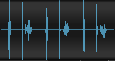
This is an example of a mild mitral stenosis murmur which is most commonly due to rheumatic heart disease.
The first heart sound is increased in intensity due to mild thickening of the mitral valve leaflets.
The second heart sound is normal and unsplit
Systole is silent.
There is an opening snap 100 milliseconds into diastole. As mitral stenosis becomes more severe, the opening snap will occur earlier in diastole.
The opening snap is followed by a diamond shaped low-frequency murmur. Use the bell of the stethoscope to hear this murmur.
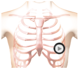
In the animation you can see the turbulent blood flow from the left atrium into the left ventricle. You can see the minimally thickened mitral valve leaflets and the minimally enlarged left atrium. The excursion of the mitral valve leaflets is minimally decreased.
Mitral Stenosis - Mild Audio
Technique
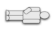
The patient's position should be supine left side down.
Auscultation Tips for Mitral Stenosis - Mild
S1:Increased intensityDiastole:Opening snap followed by diamond shaped, low-pitched murmur
Sound Wave
Mitral Stenosis - Mild Video
Review the cardiac animation. You can see the turbulent blood flow from the left atrium into the left ventricle. Observe the minimally thickened mitral valve leaflets and the minimally enlarged left atrium. The excursion of the mitral valve leaflets is minimally decreased.
Authors and Sources
Authors and Reviewers
- ECG heart rhythm modules: Thomas O'Brien.
- ECG monitor simulation developer: Steve Collmann
-
12 Lead Course: Dr. Michael Mazzini, MD.
- Spanish language ECG: Breena R. Taira, MD, MPH
- Medical review: Dr. Jonathan Keroes, MD
- Medical review: Dr. Pedro Azevedo, MD, Cardiology
- Last Update: 11/8/2021
Sources
-
Electrocardiography for Healthcare Professionals, 6th Edition
Kathryn Booth and Thomas O'Brien
ISBN10: 1265013470, ISBN13: 9781265013479
McGraw Hill, 2023 -
Rapid Interpretation of EKG's, Sixth Edition
Dale Dublin
Cover Publishing Company -
EKG Reference Guide
EKG.Academy -
12 Lead EKG for Nurses: Simple Steps to Interpret Rhythms, Arrhythmias, Blocks, Hypertrophy, Infarcts, & Cardiac Drugs
Aaron Reed
Create Space Independent Publishing -
Heart Sounds and Murmurs: A Practical Guide with Audio CD-ROM 3rd Edition
Elsevier-Health Sciences Division
Barbara A. Erickson, PhD, RN, CCRN -
The Virtual Cardiac Patient: A Multimedia Guide to Heart Sounds, Murmurs, EKG
Jonathan Keroes, David Lieberman
Publisher: Lippincott Williams & Wilkin)
ISBN-10: 0781784425; ISBN-13: 978-0781784429 - Project Semilla, UCLA Emergency Medicine, EKG Training Breena R. Taira, MD, MPH
-
ECG Reference Guide
PracticalClinicalSkills.com
Mitral Stenosis - Mild | Auscultation Cheat Sheet with Sounds & Video | #96