Cardiac Monitoring EKG Module
Cardiac Monitoring
&
Einthoven's Triangle
Thomas E. O'Brien
AS CCT CRAT RMA CCMA
Lessons
Lesson #1: Introduction 324
Part 1
Cardiac monitoring is commonly performed in many areas in critical care. Applying a cardiac monitor is a simple task, but the circumstances may be very different from prehospital to the emergency department to surgery, including pre and post-operatively and the myriad of intensive care units and telemetry.
Part 2
The important thing to remember is that the patient has a monitor applied for a specific reason. Remain diligent in your observation and report any changes noted during "monitoring".
A cardiac monitor is a tool to be used to assist with assessment of the patient. Remember to always treat the patient and not the "monitor".
Part 3
An unresponsive patient with what appears to be Normal Sinus Rhythm may in fact be clinically dead. They may have something called "Pulseless Electrical Activity".
Pay strict attention to your patient's vital signs and level of consciousness during monitoring.
Lesson #2: Introduction (Cont)
Part 4 (Cont)
Treat the patient, not the monitor.
Loose, or dried out sensors (electrodes) or broken cables may cause a tracing to appear much the same as one of these life-threatening dysrhythmias.
Part 5
What appears to be Ventricular Tachycardia might occur because the patient is brushing their teeth. This is referred to as "toothbrush tachycardia".
- This is caused by the repetitive body movement when brushing your teeth.
- This is not an actual cardiac event.
- Remember if you have an unresponsive patient, call a "code blue" (follow your facility's protocol) and begin emergency procedures.
Part 6
A variety of manufacturers offer a wide variety of cardiac monitors. A majority will have three or five cables. We will focus on the most common type today, the three-cable device.
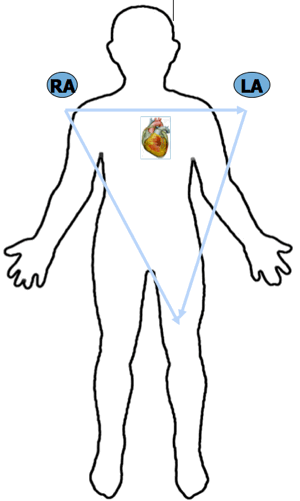
Lesson #3: Sensor Placement
Triangle
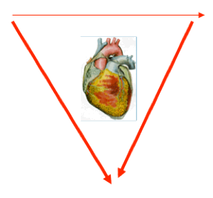
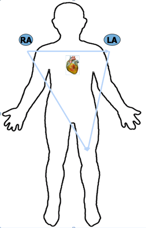
The three sensors applied may be placed in a few different locations, but ultimately they form a "triangle" around the heart.
This often referred to as Einthoven's Triangle.
Locations
- One sensor is placed on either the right wrist, right deltoid or upper outer aspect of the chest, just inside the right shoulder.
- The next sensor is placed on either the left wrist, left deltoid or upper outer aspect of the chest, just inside the left shoulder.
- Finally, this last sensor is placed at either the inner left lower leg (just above the ankle) or at the left lateral costal margin of the ribs.
Lesson #4: Views 324
Introduction
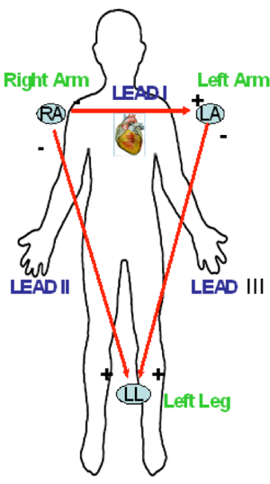
Leads I, II, & III are referred to as "bipolar leads". This is due to having two sensors on the skin surface making the positive and negative pole for this particular view.
When monitoring patients in one of these views, the sensor not involved in the "lead circuit" will become the ground.
Which Leads?
Einthoven's Triangle is formed by which three leads?
Answer
Einthoven's Triangle is formed by which three leads?
- Right Shoulder (arm)
- Left Shoulder (arm)
- Left Leg
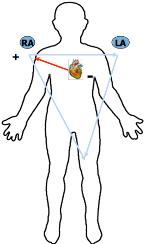
Lesson #5: Einthoven Triangle
Introduction
By use of three sensors, one each placed on the right arm, left arm and left leg, we are able to obtain six different views.
The first three views I will review are called Standard Leads I, II and III These views are referred to as "bipolar leads" due to two sensors utilized on the skin surface (one is positive and the other is negative – just like a battery) to complete the circuit.
The other three views are aVR, aVL and aVF. These will be discussed shortly.
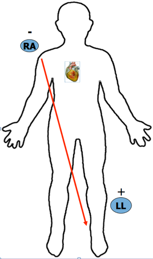
One last comment, direct views of the heart will always be from the position of the positive pole i.e. Lead II and aVF positive pole is the left leg. These would be "inferior" views.
Overview
Einthoven's Triangle
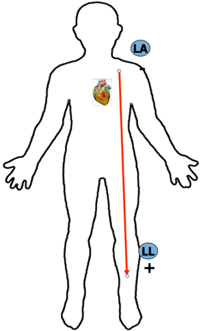
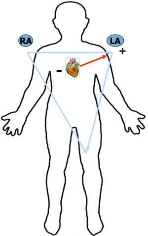
Lesson #6: Lead II
Where is the ground?
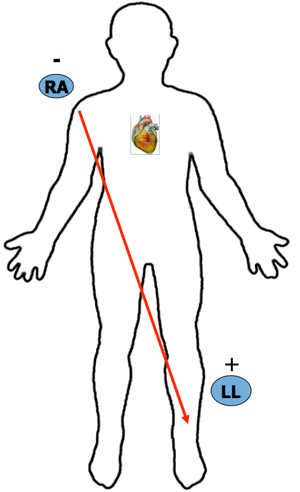
Answer
Left Arm (shoulder)
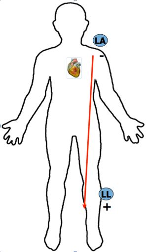
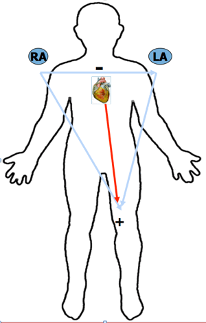
Lesson #7: Lead III
Where is the ground?
Answer
Right Arm (shoulder)
Lesson #8: Augmented Limb Leads
Discussion
The heart is the "negative" pole or terminus for all augmented leads.
The sensor on the skin surface is the positive (+) pole.
Augmented leads are "unipolar" due to one sensor being on the skin surface.
The flow of electricity is measured from the heart to either the:
- right arm (aVR)
- left arm (aVL)
- left leg or foot (aVF)
Lesson #9: aVR Illustrations
aVR Question
aVR views the heart from which direction?

aVR Answer
aVR views the heart from which direction? Right Arm
Lesson #10: aVL Illustrations
aVL Question
aVL views the heart from which direction?
aVL Answer
aVL views the heart from which direction? Left Arm
Lesson #11: aVF Illustrations
aVF Question
aVF views the heart from which direction?
aVF Answer
aVF views the heart from which direction? Left Foot (Leg)
Lesson #13: Quiz Test Questions 324
Question #1
Lead I is formed by which two locations?
B. Left arm and left leg
C. Right arm and left leg
D. Left arm and right arm
Question #2
The ground for Lead III is located on the
B. Left leg
C. Right arm
D. Left arm
Question #3
Lead II is formed by which two locations?
B. Left arm and left leg
C. Right arm and left leg
D. Left arm and right arm
Authors and Reviewers
- EKG heart rhythm modules: Thomas O'Brien
-
Medical review: Dr. Jonathan Keroes, MD
- Medical review: Dr. Pedro Azevedo, MD, Cardiology
-
Last Update: 11/8/2021
Sources
-
Electrocardiography for Healthcare Professionals, 6th Edition
Kathryn Booth and Thomas O'Brien
ISBN10: 1265013470, ISBN13: 9781265013479
McGraw Hill, 2023 -
Rapid Interpretation of EKG's, Sixth Edition
Dale Dublin
Cover Publishing Company -
EKG Reference Guide
EKG.Academy -
12 Lead EKG for Nurses: Simple Steps to Interpret Rhythms, Arrhythmias, Blocks, Hypertrophy, Infarcts, & Cardiac Drugs
Aaron Reed
Create Space Independent Publishing -
The Virtual Cardiac Patient: A Multimedia Guide to Heart Sounds, Murmurs, EKG
Jonathan Keroes, David Lieberman
Publisher: Lippincott Williams & Wilkin)
ISBN-10: 0781784425; ISBN-13: 978-0781784429 -
ECG Reference Guide
PracticalClinicalSkills.com