Lessons and Practice Strips
ECG Practice Strips
Lesson #1: Rhythm Analysis 316
Intro
The 5 steps of rhythm analysis should be followed when analyzing any rhythm strip. Analyze tracings in the following sequence.
- Rhythm Regularity
- Heart Rate
- P wave morphology
- P R interval or PRi
- QRS complex duration and morphology
Step 1
Rhythm Regularity
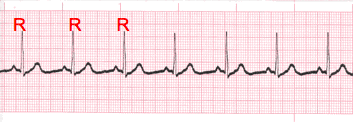
- Carefully measure from the tip of one R wave to the next, from the beginning to the end of the tracing.
- A rhythm is considered “regular or constant” when the distance apart is either the same or varies by 1 ½ small boxes or less from one R wave to the next R wave.
Step 2
Heart Rate Regular (Constant) Rhythms
- The heart rate determination technique used will be the 1500 technique.
- Starting at the beginning of the tracing through the end, measure from one R wave to the next R wave (ventricular assessment), then P wave to P wave (atrial assessment), then count the number of small boxes between each and divide that number into 1500. This technique will give you the most accurate heart rate when analyzing regular heart rhythms. You may include ½ of a small box i.e. 1500/37.5 = 40 bpm (don’t forget to round up or down if a portion of a beat is included in the answer).
Step 2-2
Heart Rate - Irregular Rhythms
- If the rhythm varies by two small boxes or more, the rhythm is considered “irregular”.
- The heart rate determination technique used for irregular rhythms will be the “six-second technique”.
- Simply count the number of cardiac complexes in six seconds and multiply by ten.
Step 3
P wave Morphology (shape)
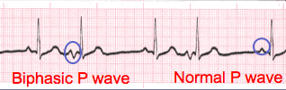
- Lead II is most commonly referenced in cardiac monitoring
- In this training module, lead two will specifically be referenced unless otherwise specified.
Step 4
PR interval (PRi)
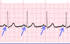 Constant PR Interval
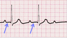 Variable PR Interval
- Measurement of the PR interval reflects the amount of time from the beginning of atrial depolarization to the beginning of ventricular depolarization.
- Plainly stated, this measurement is from the beginning of the P wave to the beginning of the QRS complex.
- The normal range for PR interval is: 0.12 – 0.20 seconds (3 to 5 small boxes)
- It is important that you measure each PR interval on the rhythm strip.
- Some tracings do not have the same PRi measurement from one cardiac complex to the next. Sometimes there is a prolonging pattern, sometimes not.
- If the PR intervals are variable, report them as variable, but note if a pattern is present or not.
Step 5
QRS complex
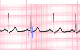
- QRS represents ventricular depolarization.
- It is very important to analyze each QRS complex on the tracing and report the duration measurement and describe the shape (including any changes in shape).
- As discussed in step 3, when referring to P waves, remember changes in the shape of the waveform can indicate the locus of stimulation has changed or a different conduction pathway was followed. It is no different when analyzing the QRS complex. The difference is that in step 3, we were looking at atrial activity. Now we are looking at ventricular activity.
- Measure from the beginning to the end of ventricular depolarization.
- The normal duration of the QRS complex is: 0.06 – 0.10 second
Close
- The previous slides presented the five-steps of rhythm analysis. These five steps must be followed regardless of how simple of complex the tracing is you are reviewing.
- The information gathered in these steps are telling a story.
- The title of that story is the interpretation.
Lesson #2: Heart Block Dysrhythmias
Introduction to Heart Blocks Part 1
- Heart Block ECG dysrhythmias occur for a variety reasons. It may be congenital as is often the case in First Degree Heart Block. They may occur secondary to medications or the result of transient illness or disease which results in tissue death affecting a portion of the conduction system.
- Each heart block (AV Block) ECG has at least one distinctive feature making it unique when comparing them with other heart blocks and dysrhythmias in other categories.
- We will focus on those “unique” features during this presentation.
Part 2
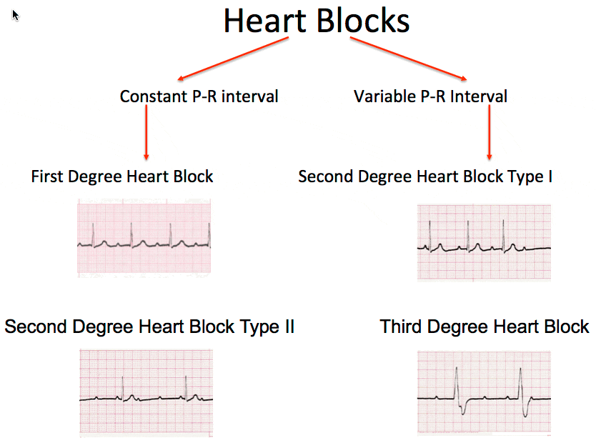
- Sometimes organizing heart block EKGs can help you to separate one heart block from another.
- On the next slide we have organized heart blocks by constant vs. variable P-R interval.
Heart Blocks Chart
Lesson #3: First Degree Heart Block
Description
- First Degree Heart Block will look like a typical sinus rhythm with one distinguishing feature.
- The P-R interval will be constant throughout the tracing and measure greater than 0.20 seconds.
- Rate, regularity, P wave morphology and QRS duration and morphology will be unaffected.
- NOTE: The rate will be that of the underlying rhythm. If the rate is “normal”, it will be 60 – 100 bpm. If it is bradycardia, the rate will be less than 60 bpm.
- First Degree Heart Block is the most common heart block.
- People are born with it every day. They will likely live a long and healthy life and die from some other malady.
- Patients who develop a heart block during an MI bear close observation. This must be reported to the licensed healthcare practitioner immediately.
Example
Notice the following: the only abnormality when analyzing this tracing is the abnormal duration of the P-R interval.
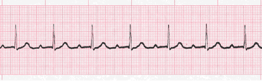
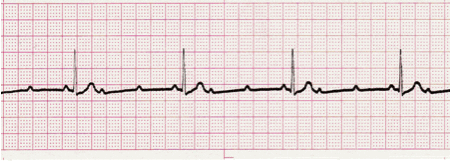
Practice Strip
Analyze this tracing using the five steps of rhythm analysis.
Show Answer
- Rhythm: Regular
- Rate: 68
- P Wave: Upright
- PR interval: 0.28 sec
- QRS: 0.08 sec
- Interpretation: First Degree Heart Block
Lesson #4: Second Degree Heart Block Type I
Description
- Also known as Wenckebach Phenomenon; second degree heart block ECG dysrhythmia is typically stable and often temporary with the patient remaining asymptomatic as long as the ventricular response remains within the “normal” range.
- The unique feature (hallmark) of this dysrhythmia is the presence of a prolonging P-R interval from one cardiac complex to the next, until it reaches a point where the QRS complex is non-conducted ( blocked or more simply missing). Then the pattern starts over again.
Example
- In this dysrhythmia, if the third QRS complex is dropped/blocked, then it will always be the third complex that is blocked before re-setting in a repetitious pattern.
- It is important to note the following:
- The P – P intervals are regular and the R to R intervals are irregular.
- here are more P waves than QRS complexes. Report the rate of each separately.
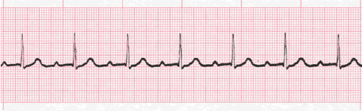
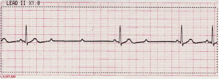
Practice Strip
Analyze this tracing using the five steps of rhythm analysis.
Show Answer
- Rhythm: Atria – Regular, Ventricles - Irregular
- Rate: Atria – 56, Ventricles - 40
- P Wave: Upright
- PR interval: Variable, progressive
- QRS: 0.10 sec
- Interpretation: Second Degree Heart Block Type I
Lesson #5: Second Degree Heart Block Type II
Description
- The hallmark of this dysrhythmia is a constant P-R interval with missing QRS complexes.
- This dysrhythmia may present in a couple of different ways.
- A. QRS complexes occurring in a
specific pattern in a ratio with the P
waves. This is often referred to as
2:1 or 3:1 block depending upon
the ratio of P waves to each QRS
complex.
- B. QRS complexes occur in a more
unstable, unpredictable manner.
-
Either presentation requires immediate reporting due to its potential for conversion to Third Degree (Complete) Heart Block.
A.
B.
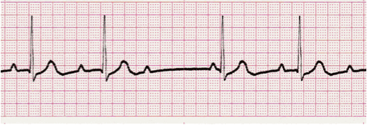
Practice Strip
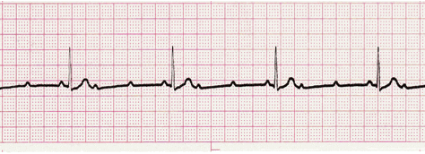
Analyze this tracing using the five steps of rhythm analysis.
Show Answer
- Rhythm: Atria and Ventricles - Regular
- Rate: Atria – 125, Ventricles - 41
- P Wave: Upright
- PR interval: 0.14 sec
- QRS: 0.06
- Interpretation: Second Degree Heart Block Type II, 3:1
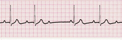
Practice Strip #2
Analyze this tracing using the five steps of rhythm analysis.
Show Answer
- Rhythm: A – Regular, V - Irregular
- Rate: Atria – 68, Ventricles - 40
- P Wave: Upright
- PR interval: 0.16 sec
- QRS: 0.08
- Interpretation: Second Degree Heart Block Type II
Lesson #6: Third Degree Heart Block
Description Part 1
- Third Degree Heart Block is also known as “Complete Heart Block”.
- This name more accurately describes the electrical event or problem occurring within the heart.
- As a result of disease or tissue death, there is a blockage preventing electrical impulses within the atria from entering the ventricular conduction system.
- The outcome of 3rd degree heart block EKG are two independently functioning pacemakers within the heart (typically one is supraventricular, the other is ventricular).
- Essentially, the atria and ventricles are electrically separated (dissociated) from one another.
Description Part 2
- What will be seen are regularly occurring P waves and QRS complexes, but at two distinctly different rates.
- The QRS complexes may occur as a result of impulses coming from the His bundle or the Purkinje network.
- Morphology and rate will often provide clues regarding the locus of ventricular impulse stimulation. A more narrow appearing QRS with a rate greater than 40 generally would indicate the impulse formation is coming from the His bundle. A wide, bizarre QRS with a rate of 40 or less indicates the impulses are originating in the Purkinje network.
- Complete heart block presents with Regular P to P and R to R intervals and a variable P-R interval.
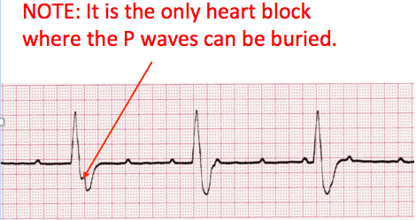
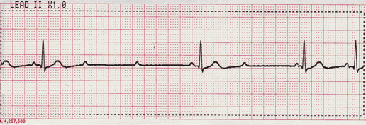
ECG Practice Strip
Analyze this tracing using the five steps of rhythm analysis.
Show Answer
- Rhythm: Atria and Ventricles - Regular
- Rate: Atria – 94, Ventricles - 34
- P Wave: Upright (some buried)
- PR interval: Variable
- QRS: 0.14 sec
- Interpretation: Third Degree (Complete) Heart Block
Lesson #7: Quiz Test Questions 316