Tetralogy of Fallot Lesson
Where to Auscultate
The patient was supine during auscultation.
Description
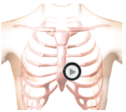
This lesson presents Tetralogy of Fallot, which is a congenital condition often called Blue Baby Syndrome. It is characterized by four abnormalities: - pulmonic stenosis - increased thickening of the right ventricle - a ventricular septal defect - overriding aorta S1 and S2 are normal and unsplit. There is an aortic ejection click in systole. A diamond shaped murmur following the click and ending well before the second heart sound. In the cardiac animation video, observe the turbulent flow from the right ventricle into the pulmonary artery across the stenotic pulmonic valve and turbulent flow from the left ventricle to the right ventricle (the ventricular septal defect). The right ventricular wall is thickened. If you listen at the tricuspid position, you are hearing the ventricular septal defect. If you listen at the pulmonic area, you are hearing the pulmonic stenosis. Both create diamond-shaped systolic murmurs.
Phonocardiogram
Anatomy
Tetralogy of Fallot
Authors and Sources
Authors and Reviewers
-
Heart sounds by Dr. Jonathan Keroes, MD and David Lieberman, Developer, Virtual Cardiac Patient.
- Lung sounds by Diane Wrigley, PA
- Respiratory cases: William French
-
David Lieberman, Audio Engineering
-
Heart sounds mentorship by >W. Proctor Harvey, MD
- Special thanks for the medical mentorship of Dr. Raymond Murphy
- Reviewed by Dr. Barbara Erickson, PhD, RN, CCRN.
-
Last Update: 11/10/2021
Sources
-
Heart and Lung Sounds Reference Library
Diane S. Wrigley
Publisher: PESI -
Impact Patient Care: Key Physical Assessment Strategies and the Underlying Pathophysiology
Diane S Wrigley & Rosale Lobo - Practical Clinical Skills: Lung Sounds
- PESI Faculty - Diane S Wrigley
-
Case Profiles in Respiratory Care 3rd Ed, 2019
William A.French
Published by Delmar Cengage -
Essential Lung Sounds
by William A. French
Published by Cengage Learning, 2011 -
Understanding Lung Sounds
Steven Lehrer, MD
-
Clinical Heart Disease
W Proctor Harvey, MD
Clinical Heart Disease
Laennec Publishing; 1st edition (January 1, 2009) -
Heart and Lung Sounds Reference Guide
PracticalClinicalSkills.com