Coarctation of the Aorta 698 Lesson
Where to Auscultate
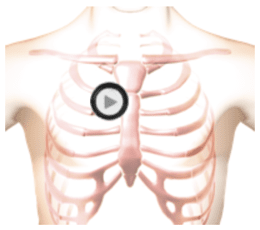

The patient was sitting leaning forward during auscultation.
Description
This lesson presents coarctation of the aorta, a congenital abnormality. S1 is normal, and the S2 sound is intensified. A diamond-shaped murmur spans most of systole while a high-pitched decrescendo murmur occurs in the first half of diastole.
In the cardiac anatomy video, observe a constriction in the descending aorta, which is responsible for the systolic murmur. There is regurgitant blood flowing from the aorta into the left ventricle producing the diastolic murmur. The left ventricle wall thickness is increased due to aortic pressure elevation caused by the aortic coarctation.
Phonocardiogram
Anatomy
Coarctation of the Aorta 698
Play the cardiac movie and observe a constriction in the descending aorta which is responsible for the systolic murmur.
There is regurgitant flow from the aorta into the left ventricle which causes the diastolic murmur.
The left ventricle wall thickness is increased due to aortic pressure elevation caused by the aortic coarctation.
Authors and Sources
Authors and Reviewers
-
Heart sounds by Dr. Jonathan Keroes, MD and David Lieberman, Developer, Virtual Cardiac Patient.
- Lung sounds by Diane Wrigley, PA
- Respiratory cases: William French
-
David Lieberman, Audio Engineering
-
Heart sounds mentorship by >W. Proctor Harvey, MD
- Special thanks for the medical mentorship of Dr. Raymond Murphy
- Reviewed by Dr. Barbara Erickson, PhD, RN, CCRN.
-
Last Update: 11/10/2021
Sources
-
Heart and Lung Sounds Reference Library
Diane S. Wrigley
Publisher: PESI -
Impact Patient Care: Key Physical Assessment Strategies and the Underlying Pathophysiology
Diane S Wrigley & Rosale Lobo - Practical Clinical Skills: Lung Sounds
- PESI Faculty - Diane S Wrigley
-
Case Profiles in Respiratory Care 3rd Ed, 2019
William A.French
Published by Delmar Cengage -
Essential Lung Sounds
by William A. French
Published by Cengage Learning, 2011 -
Understanding Lung Sounds
Steven Lehrer, MD
-
Clinical Heart Disease
W Proctor Harvey, MD
Clinical Heart Disease
Laennec Publishing; 1st edition (January 1, 2009) -
Heart and Lung Sounds Reference Guide
PracticalClinicalSkills.com