Normal Heart Sounds Lesson
Where to Auscultate
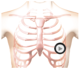
The patient was supine during auscultation.
Description
The closure of the mitral and tricuspid valves creates the first heart sound. The mitral valve usually closes first, immediately followed by the tricuspid valve. Closure of the aortic and pulmonic valves creates the second heart sound.
This recording is a normal first and second heart sound with a rate of sixty beats per minute. The stethoscope's diaphragm is positioned at the Apex (mitral valve area). Usually, the first heart sound is somewhat louder than the second heart sound when auscultating at the Apex. Sound intensity will vary with the chestpiece's position as well as with the patient's anatomy.
In addition to sound intensity, the first heart sound (S1) can be identified by its timing. At moderate heartbeat rates, the first heart sound follows the longer pause.
Phonocardiogram
Anatomy
Normal Heart Sounds
Authors and Sources
Authors and Reviewers
-
Heart sounds by Dr. Jonathan Keroes, MD and David Lieberman, Developer, Virtual Cardiac Patient.
- Lung sounds by Diane Wrigley, PA
- Respiratory cases: William French
-
David Lieberman, Audio Engineering
-
Heart sounds mentorship by >W. Proctor Harvey, MD
- Special thanks for the medical mentorship of Dr. Raymond Murphy
- Reviewed by Dr. Barbara Erickson, PhD, RN, CCRN.
-
Last Update: 11/10/2021
Sources
-
Heart and Lung Sounds Reference Library
Diane S. Wrigley
Publisher: PESI -
Impact Patient Care: Key Physical Assessment Strategies and the Underlying Pathophysiology
Diane S Wrigley & Rosale Lobo - Practical Clinical Skills: Lung Sounds
- PESI Faculty - Diane S Wrigley
-
Case Profiles in Respiratory Care 3rd Ed, 2019
William A.French
Published by Delmar Cengage -
Essential Lung Sounds
by William A. French
Published by Cengage Learning, 2011 -
Understanding Lung Sounds
Steven Lehrer, MD
-
Clinical Heart Disease
W Proctor Harvey, MD
Clinical Heart Disease
Laennec Publishing; 1st edition (January 1, 2009) -
Heart and Lung Sounds Reference Guide
PracticalClinicalSkills.com