Ventricular Septal Defect | Auscultation #62 | Lesson with Audio
Where to Auscultate
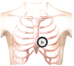
The patient was supine during auscultation.
Description
This is an example of ventricular septal defect as heard at the tricuspid position. Ventricular Septal Defect is a congenital condition associated with abnormal blood flow between the left ventricle and the right ventricle. During fetal development a wall develops creating a right and left ventricle. In a percentage of individuals a defect in the wall persists allowing blood flow from the left ventricle into the right ventricle. This condition is known as a ventricular septal defect. The first heart sound is normal. The second heart sound is unsplit. There is a third heart sound followed by a short diamond shaped diastolic murmur. A medium pitched murmur fills all of systole. In the anatomy video you see an enlarged right ventricle and an enlarged left atrium. You see turbulent blood flow from the left ventricle into the right ventricle through the up portion of the septum (the systolic murmur). There is further turbulent flow into the left ventricle from the left atrium causing the diastolic murmur. This is caused by VSD induced increased blood flow across the mitral valve.Phonocardiogram
Anatomy
Ventricular Septal Defect
Observe an enlarged right ventricle and an enlarged left atrium in the cardiac animation.
Authors and Sources
Authors and Reviewers
-
Heart sounds by Dr. Jonathan Keroes, MD and David Lieberman, Developer, Virtual Cardiac Patient.
- Lung sounds by Diane Wrigley, PA
- Respiratory cases: William French
-
David Lieberman, Audio Engineering
-
Heart sounds mentorship by >W. Proctor Harvey, MD
- Special thanks for the medical mentorship of Dr. Raymond Murphy
- Reviewed by Dr. Barbara Erickson, PhD, RN, CCRN.
-
Last Update: 11/10/2021
Sources
-
Heart and Lung Sounds Reference Library
Diane S. Wrigley
Publisher: PESI -
Impact Patient Care: Key Physical Assessment Strategies and the Underlying Pathophysiology
Diane S Wrigley & Rosale Lobo - Practical Clinical Skills: Lung Sounds
- PESI Faculty - Diane S Wrigley
-
Case Profiles in Respiratory Care 3rd Ed, 2019
William A.French
Published by Delmar Cengage -
Essential Lung Sounds
by William A. French
Published by Cengage Learning, 2011 -
Understanding Lung Sounds
Steven Lehrer, MD
-
Clinical Heart Disease
W Proctor Harvey, MD
Clinical Heart Disease
Laennec Publishing; 1st edition (January 1, 2009) -
Heart and Lung Sounds Reference Guide
PracticalClinicalSkills.com