
Introduction
This complex murmurs module includes six lessons. We provide a textual description, audio recording, dynamic waveform video, and a cardiac animation for each lesson. Optionally, a quiz can be taken to measure comprehension and listening skills. Users who have selected our Auscultation-Basics, Essentials or Advanced plans can print achievement certificates and view their progress and scores using our personalized dashboard.
- Recognize murmurs that include multiple abnormal sounds
- Identify pan-systolic murmurs
- Identify key sound attributes of complex murmurs
Overview
Complex murmurs are sustained sounds occurring in systole, diastole or spanning both systole and diastole. These murmurs are considered abnormal. Complex murmurs causes include valvular problems, enlarged atria or enlarged ventricles.
When auscultating, take note of these murmur attributes:
- Location
- Timing: early, mid, late or pansystolic
- Loudness and its pattern during systole
- Pitch
- Quality (harsh, blowing or musical)
- Murmur changes during the respiratory cycle
Sound Patterns
As an introduction, we present two sound patterns that illustrate complex murmurs. We will cover these in the lessons that follow.
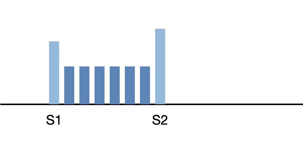
This sound diagram illustrates a holosystolic murmur. The murmur begins with the first heart sound and continues until the second heart sound, with a fairly uniform intensity.
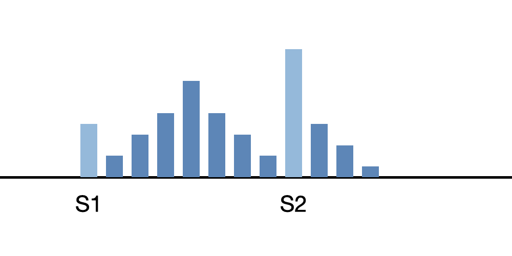
This sound diagram illustrates a complex systolic/diastolic murmur. We have dedicated a full lesson to this type of murmur.
The lessons in this course present several types of abnormal, complex murmurs, including their attributes and clinical correlations.
Start Complex Murmurs Course
Complex Murmurs Lessons
Severe Mitral Regurgitation
This lesson presents severe mitral regurgitation. The first heart sound is normal while the second heart sound is widely split. A third heart sound gallop follows S2. A loud, rectangular, pansystolic murmur is present. In addition, a brief, rumbling, diamond-shaped diastolic murmur immediately after the third heart sound.
In the cardiac anatomy video, you can see the enlarged left atrium and left ventricle. Observe the turbulent blood flow from the left ventricle into the left atrium. This is the systolic murmur. Also notice the brief turbulent blood flow from the left atrium to the left ventricle during diastole. This is caused by too much blood in the left atrium which forces blood back into the ventricle during diastole producing the flow rumble. Severe mitral regurgitation is often caused by degeneration of the mitral valve leaflets.
Severe Mitral Regurgitation Lesson
Severe Tricuspid Regurgitation
This recording is an example of severe tricuspid regurgitation. The first heart sound is normal and S2 is unsplit. A loud, rectangular, pansystolic murmur is present as well as a brief, rumbling, diamond-shaped diastolic murmur.
In the cardiac animation video, observe the enlarged right atrium and right ventricle. Notice the turbulent blood flow from the right ventricle into the right atrium. This is the systolic murmur. Observe the brief turbulent blood flow from the right atrium to the right ventricle in diastole. This is caused by too much blood in the right atrium which forces blood back into the ventricle during diastole producing the flow rumble.
To differentiate tricuspid regurgitation from mitral regurgitation, the maximum intensity of the tricuspid murmur is heard at the left lower sternal border. In addition, the murmur intensity increases with inspiration.
Severe Tricuspid Regurgitation Lesson
Severe Mitral Stenosis and Mild Mitral Regurgitation
This lesson presents severe mitral stenosis combined with mild mitral regurgitation in a patient with rheumatic heart disease. The first heart sound is slightly louder than normal. The second heart sound is unsplit. There is an opening snap fifty milliseconds after the second heart sound.
This murmur includes a low-frequency murmur filling all of diastole. The first two-thirds of the murmur is diamond-shaped and the last third is a crescendo.
There is a rectangular medium frequency murmur which fills the first half of systole.
In the cardiac animation video, observe an enlarged left atrium and thickened mitral valve leaflets which barely moved. The turbulent blood flow represents the systolic and diastolic murmurs.
Severe Mitral Stenosis and Mild Mitral Regurgitation Lesson
Mild Aortic Stenosis Moderate and Regurgitation
This lesson discusses moderate aortic stenosis combined with mild aortic regurgitation in a patient with rheumatic heart disease.
S1 is normal, and S2 is unsplit. An aortic ejection click occurs in systole followed by a diamond-shaped systolic murmur. A high-pitched decrescendo murmur fills the first two thirds of diastole.
In the cardiac animation video, you can see a thickened left ventricle and thickened aortic valve leaflets. Moderate turbulent blood flows across the aortic valve in systole and a mild regurgitant turbulent flow into the left ventricle occurs in diastole. These turbulent blood flows cause the systolic and diastolic murmurs.
Mild Aortic Stenosis Moderate and Regurgitation Lesson
Mitral Regurgitation and Aortic Regurgitation
This is an example of acute pericarditis Murmurs are generated by turbulent blood flow across incompetent or stenotic valves. In contrast, a pericardial friction rub is produced by the rubbing together of two surfaces of the pericardial sack. The pericardial friction rub has three parts; a systolic component, an early diastolic component and a late diastolic component. The first and second heart sounds are obscured by the rubbing sounds.
This murmur is auscultated at Erb's Point.
In the cardiac animation video, observe the yellow fluid accumulation around the heart caused by an inflamed pericardial sack.
Mitral Regurgitation and Aortic Regurgitation Lesson
Acute Pericarditis
This is an example of acute pericarditis Murmurs are generated by turbulent blood flow across incompetent or stenotic valves. In contrast, a pericardial friction rub is produced by the rubbing together of two surfaces of the pericardial sack. The pericardial friction rub has three parts; a systolic component, an early diastolic component and a late diastolic component. The first and second heart sounds are obscured by the rubbing sounds.
This murmur is auscultated at Erb's Point.
In the cardiac animation video, observe the yellow fluid accumulation around the heart caused by an inflamed pericardial sack.
Course Quiz
After completing all lessons in a course, a quiz becomes available. If the user successfully completes a quiz, results are saved to the user's dashboard and a certificate can be printed.
Reference Guide
For subscribers, we provide a comprehensive heart sounds and murmurs reference guide. For each abnormality, one or more sound recordings are available along with text, phonocardiogram and cardiac animation.Course Completion
Registered users can earn a certificate of achievement for this module by reading all content and then earning a passing score on this module's quiz.
Completed modules and related scores can be viewed on the dashboard.
Authors and Sources
Authors and Reviewers
-
Heart sounds by Dr. Jonathan Keroes, MD and David Lieberman, Developer, Virtual Cardiac Patient.
- Reviewed by Dr. Barbara Erickson, PhD, RN, CCRN.
-
Last Update: 11/9/2021
Sources
-
Heart Sounds and Murmurs Across the Lifespan (with CD)
Dr Barbara Ann Erickson
Publisher: Mosby
ISBN-10: 0323020453; ISBN-13: 978-0323020459 -
Heart Sounds and Murmurs: A Practical Guide with Audio CD-ROM 3rd Edition
Elsevier-Health Sciences Division
Barbara A. Erickson, PhD, RN, CCRN - How to measure blood pressure using a manual monitor
Mayo Foundation for Medical Education and Research (MFMER) -
Heart and Lung Sounds Reference Guide
PracticalClinicalSkills.com -
Manual Blood Pressure Measurement
Vital Sign Measurement Across the Lifespan